What Are Lymph Nodes, and When Does Swelling Require Attention?
Most people have heard of lymph nodes but couldn’t tell you exactly where they are or what they do. They’re part of the immune system, small structures located in clusters throughout the body that filter lymph fluid and house immune cells ready to respond to infections and other threats. Normally, you can’t feel them at all. When they enlarge to the point of being noticeable, that’s the immune system flagging something. The question is always: flagging what?
Star of Texas Veterinary Hospital is a Fear Free certified practice in Austin, which means the evaluation process stays as comfortable as possible for your pet even when the findings are concerning. We provide checkups to your pet in the manner that works best for them- whether that’s in our own backyard, with pre-visit anti-anxiety medications, in your lap, or through extended appointments where we move at your pet’s pace. Our diagnostic services include thorough workups for lymph node abnormalities, with clear communication throughout. Contact us if you’ve found something that doesn’t belong or noticed changes you’re worried about.
What Do Lymph Nodes Do and Where Are They Located?
Lymph nodes are immune checkpoints. They filter lymph fluid (the clear fluid that drains from tissues and circulates through the lymphatic system), capture pathogens before they spread further, and house immune cells that coordinate the response when something harmful is detected. Most of the time they work invisibly. Enlargement is a sign that the immune system has detected something and is mounting a response.
The accessible lymph node locations you can check at home:
- Submandibular (under the jaw, on either side of the throat)
- Prescapular (in front of the shoulders, where the neck meets the chest)
- Axillary (in the armpit area, behind the front legs)
- Inguinal (in the groin, where the rear legs meet the body)
- Popliteal (behind each knee, on the back of the rear leg)
Internal lymph nodes (in the chest, abdomen, and along major vessels) aren’t palpable from outside. Imaging is needed to assess them. The visual lymph node chart frameworks are useful references for identifying the locations during home checks.
Lymph node palpation is a standard part of every preventive care visit at Star of Texas. We’re happy to show you where and what to feel for- just ask our team.
What Causes Lymph Nodes to Swell in Pets
The pattern of involvement provides meaningful diagnostic information before any testing begins. A single enlarged node near a wound suggests a local infection. Multiple nodes enlarged across different body regions suggests a systemic process. Firm, fixed nodes that aren’t tender raise concern for cancer. Soft, painful nodes more often suggest active infection.
The major categories of causes:
Infections and Reactive Lymph Node Swelling in Pets
Bacterial, viral, fungal, and parasitic infections are among the most common causes of lymphadenopathy.
Bacterial and tick-borne infections: Lyme disease can cause regional lymph node enlargement. Ehrlichia and anaplasma often produce both lymph node swelling and other systemic signs. Leptospirosis involves lymph nodes alongside kidney and liver effects. Mycobacteriosis (less common but significant) causes characteristic granulomatous lymphadenopathy in cats.
Fungal disease often produces marked lymph node enlargement. Valley Fever is endemic in the southwestern US and causes systemic disease with lymphadenopathy. Blastomycosis is more common in the midwest and southern US. Aspergillosis and histoplasmosis round out the regional fungal causes that can affect dogs and cats.
Viral diseases in cats: Feline leukemia virus and feline immunodeficiency virus both can cause lymphadenopathy directly and through the immune dysregulation they produce. Routine testing for these viruses is part of the workup for cats with unexplained lymph node enlargement.
Parasitic causes: Toxoplasmosis in cats can produce lymphadenopathy. Intestinal parasites including giardia cause lymph node enlargement in some cases. External parasites can produce regional lymphadenopathy from chronic skin irritation and secondary infections.
Cancer as a Cause of Lymph Node Swelling in Pets
Canine lymphoma is one of the most frequent causes of sudden, dramatic lymph node swelling in dogs. The classic presentation is your dog appearing entirely well, eating normally, behaving normally, with multiple peripheral lymph nodes that you notice have suddenly enlarged. By the time other systemic signs develop, the disease has often progressed.
The numbers are sobering: an estimated 1 in 15 dogs born today will develop lymphoma during their lifetime, and the risk climbs to roughly 1 in 8 for Golden Retrievers. The encouraging counterpoint is that newer blood-based screening tests can flag lymphoma months earlier than traditional methods, often before any visible signs appear, which gives families and veterinary teams a meaningful head start on treatment planning.
Lymphoma diagnosis and subtype shapes everything that follows: treatment options, expected response, and prognosis vary substantially between subtypes. Accurate early diagnosis is essential. Early-stage disease has more treatment options and better outcomes than advanced disease.
Feline lymphoma presents differently. The most common form in cats is gastrointestinal rather than peripheral lymph node-based, with weight loss, vomiting, and diarrhea as more prominent signs than enlarged lymph nodes. Feline lymphoma still causes lymphadenopathy in some cases, particularly the multicentric form.
Other types of cancer in pets can also spread to regional lymph nodes. A skin tumor near the front leg may produce enlarged axillary nodes. A mammary tumor may produce enlarged inguinal nodes. The pattern of node involvement provides clues about where the primary disease is.
Immune-Mediated Conditions, Allergies, and Other Causes of Lymph Node Swelling
Some causes are non-infectious and non-cancerous:
- Immune-mediated hemolytic anemia can produce lymphadenopathy as part of the immune dysregulation.
- Allergies with secondary skin infections often produce regional lymph node enlargement, particularly atopic dermatitis with bacterial or yeast overgrowth. This is one of the more common causes seen in general practice.
- Vaccination reactions occasionally produce transient regional lymph node enlargement, typically resolving within a few weeks.
These causes require the same systematic workup as infectious or neoplastic causes because they’re indistinguishable on the surface. Differentiation comes from testing.
How Enlarged Lymph Nodes Are Diagnosed in Pets
Diagnosis moves systematically from physical findings to targeted testing.
What the Physical Exam Reveals About Lymph Node Swelling
A thorough lymph node exam assesses several characteristics:
- Size: Mild enlargement vs. dramatic enlargement
- Texture: Soft and fluctuant vs. firm and rubbery vs. hard
- Symmetry: Are bilateral nodes equally enlarged, or is one side more affected?
- Tenderness: Painful on palpation suggests active inflammation
- Mobility: Nodes that move freely under the skin are different from fixed nodes that may indicate invasion
- Pattern: Local (one or two nodes near a problem) vs. multicentric (multiple nodes throughout the body)
- Other findings: Skin changes, fever, weight loss, organ enlargement on abdominal palpation
The pattern shapes the urgency and direction of further testing.
Fine-Needle Aspiration and Biopsy for Lymph Node Evaluation
Fine-needle aspiration (FNA) is typically the first diagnostic step. A small needle (similar to those used for bloodwork) is inserted into the enlarged node and cells are collected for microscopic evaluation. The procedure is brief, minimally invasive, and most pets tolerate it without sedation. Our Fear Free approach makes this process as comfortable as possible.
The cytology vs biopsy distinction matters. Cytology (cell-level evaluation from FNA) often gives a definitive answer for lymphoma, infectious causes, and reactive changes. When cytology is inconclusive or when subtype information is needed (particularly for lymphoma treatment planning), surgical biopsy provides the larger tissue sample needed for definitive evaluation.
Our diagnostic and surgical capabilities support both FNA and surgical biopsies of lymph nodes, with answers often back within a few days.
Blood Work, Tick Testing, and Imaging in the Lymph Node Workup
A complete workup typically includes:
- Complete blood count (CBC) to assess for systemic infection, anemia, or abnormal blood cells suggesting cancer
- Chemistry panel to evaluate organ function and identify systemic disease
- Tick-borne disease testing (4Dx panel and additional tests as indicated by region and exposure)
- Infectious disease testing for region-specific pathogens (Valley Fever in Texas, blastomycosis in some regions, fungal cultures, etc.)
- FeLV/FIV testing for cats
- Urinalysis to assess for systemic disease
- Chest radiographs to evaluate intrathoracic lymph nodes and rule out metastatic disease
- Abdominal ultrasound to evaluate intra-abdominal lymph nodes and organ changes
When lymphoma is confirmed, staging through imaging guides treatment and prognosis. Our in-house diagnostics provide rapid results, with reference lab support for specialty testing.
What Are the Treatment Options for Swollen Lymph Nodes?
Treatment follows diagnosis. Starting treatment before the cause is confirmed leads to wrong medications and delayed resolution. The major treatment categories:
- Bacterial infections are treated with appropriate antibiotics chosen based on the organism and tissue involved
- Tick-borne diseases are treated with doxycycline or other targeted antimicrobials, typically for 28 days
- Fungal infections are treated with antifungal medications, often for months, with monitoring of liver function during treatment
- Viral infections in cats are managed supportively, with attention to secondary problems
- Parasitic causes are treated with appropriate parasiticides
- Lymphoma is treated with chemotherapy protocols selected based on subtype, with the goal of remission rather than cure for most patients
- Other cancers are treated with surgery, chemotherapy, radiation, or combinations depending on the specific cancer
- Immune-mediated conditions are treated with immunosuppression
- Allergies and skin disease are treated with appropriate dermatologic therapies plus secondary infection management
For lymphoma specifically, honest conversations about what treatment looks like for each individual pet are important. Aggressive chemotherapy protocols can produce remissions of months to years for many dogs. Less aggressive protocols can offer comfort with shorter remission durations. Some families choose palliative care from the start. Each path is valid.
How Can You Reduce the Risk of Lymph Node Problems?
Not all causes of lymphadenopathy are preventable, but many common infectious triggers are, and consistent preventive care is also the most reliable way to catch everything else early.
Preventive care including individualized vaccinations addresses leptospirosis, Lyme disease, and other vaccine-preventable causes of lymphadenopathy.
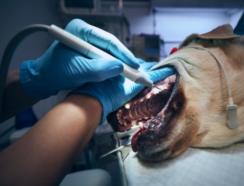
Dental care reduces the chronic bacterial load from periodontal disease that activates jaw region lymph nodes. Our comprehensive dental exams and cleanings include dental x-rays to find disease hiding under the gumline, which is where most dental abscesses and infection causing lymph node enlargement hide.
Consistent parasite prevention addresses intestinal and external parasites that can cause regional lymphadenopathy. Year-round flea and tick prevention from our pharmacy (check out our options for dogs and cats) prevents most tick-borne diseases that drive secondary lymphadenopathy.
Regular wellness exams allow lymph node abnormalities to be detected early, when they’re easier to evaluate and treat. Annual or biannual exams are the foundation; pets with prior lymph node issues may benefit from more frequent monitoring.
How Quickly to Seek Care for Swollen Lymph Nodes in Pets
Three levels of urgency:
Same-day evaluation for: rapid lymph node enlargement (over hours rather than days), multiple nodes simultaneously enlarged, lethargy or appetite loss alongside enlargement, fever, breathing difficulty, or any acute signs of systemic illness.
Within 48 hours for: a single new node that’s clearly enlarged but your pet is otherwise well, gradual enlargement noticed over days, firm or fixed feeling nodes, or any concerning physical exam finding.
Within the week for: subtle changes detected during routine home checks, mild enlargement that’s stable or slowly progressive, or general “something feels different” findings.
When uncertain which tier applies, contact us. We’re happy to help with triage decisions.
How Do You Check Your Pet’s Lymph Nodes at Home?
Monthly home checks during a relaxed petting session work well:
- Start under the jaw, feeling along both sides of the throat (submandibular nodes)
- Move to in front of the shoulders (prescapular nodes)
- Check the armpit area gently (axillary nodes)
- Check the groin where the leg meets the body (inguinal nodes)
- Behind each knee, on the back of the rear leg (popliteal nodes)
You’re not assessing exact sizes. You’re noticing change. If something feels larger or firmer than the previous month, that’s the signal to call us. Recording observations in a simple log helps track trends over time. Our Fear Free certification means even pets who dislike handling can usually tolerate brief, gentle exams done as part of regular play and affection.
Frequently Asked Questions About Swollen Lymph Nodes in Pets
Is a swollen lymph node always cancer?
No. Most lymphadenopathy in dogs and cats is reactive (from infections, allergies, or other immune activation) rather than cancerous. The diagnostic workup distinguishes the cause; we don’t assume cancer based on physical exam findings alone.
My dog’s lymph nodes feel different than I remember. Should I worry?
Possibly. Changes from a previous baseline are worth evaluation, even if you’re not sure whether the change is significant. We’d rather see a normal pet than miss a developing problem.
Can I feel my cat’s lymph nodes?
Cats have lymph nodes in the same locations as dogs, but they’re often harder to feel in healthy cats. When cat lymph nodes are clearly palpable, that’s often itself a sign of enlargement.
How quickly do enlarged lymph nodes need to be evaluated?
Sudden, dramatic enlargement (especially of multiple nodes simultaneously) warrants same-day evaluation. Gradual changes can typically be scheduled within the week. When in doubt, calling sooner is better than later.
Will I need a biopsy?
Often FNA cytology answers the question without surgical biopsy. When biopsy is needed, it’s usually because subtype information is required for treatment planning (particularly with lymphoma) or cytology was inconclusive.
Moving From Discovery to a Clear Answer
The most anxious part of finding a lump or noticing changes is not knowing what they mean. A systematic diagnostic process leads to a clear answer and a plan, and earlier evaluation means more options remain available regardless of what’s found.
Our team at Star of Texas Veterinary Hospital combines the diagnostic capabilities to evaluate lymph node abnormalities with the Fear Free care approach that keeps the process comfortable for your pet. Request an appointment and we’ll work through what’s happening and what comes next.